Leif's Anatomy Tips: MusclesMuscles 3: Assorted Tips on Origins, Insertions, and Actions |
IPHY 3415 Leif Saul |
Most recent item added on: 11 Oct 2012
General tips
- There are two kinds of facts: (1) facts that are what you'd expect based on common sense, and (2) counterintuitive or surprising facts. As you study the table of origins/insertions/actions, take note of which category a fact falls into.
- The superficial muscle reaches farther, while the deeper muscle is "trapped" so it doesn't go as far. Example: The soleus muscle originates on the proximal tibia and fibula, while the gastrocnemius, which is superficial to the soleus, not surprisingly reaches higher up than the soleus and originates on the condyles of the femur.
- The "longus" muscle inserts more distal than the "brevis" because the "longus" is a longer muscle. Example: The flexor pollicis longus inserts on the distal phalanx of the pollex; the flexor pollicis brevis inserts on the proximal phalanx of the pollex.
- Although the flexor digitorum profundus lies deep to the flexor digitorum superficialis, the profundus extends all the way to the distal phalanges, whereas the superficialis inserts on the middle phalanges. (This is accomplished by the splitting of the superficialis tendons at the middle phalanges.)
- Although the great toe is on the medial side of the foot, the leg muscles that control the great toe originate on the lateral side (on the fibula).
- In muscles that have a major/minor pair in this course, if one is superior to the other, the superior muscle is always the minor, i.e.: zygomatic minor/major, rhomboid minor/major, teres minor/major.
Examples of common-sense facts:
Examples of counterintuitive facts:
Abdomen
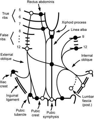
- (9/30/11) The diagram (right) schematically illustrates the origins (open dots) and insertions (filled dots) of the muscles of the abdominal wall for which you need to know OIA information. Notice that some of the lines used here to represent the external and internal obliques match the fiber direction observed on cadavers. (Note: The inferior part of internal oblique, not seen in the "windows" we cut on cadavers, actually fans out inferiorly to insert on the pubic crest as shown.) Note: only the attachments emphasized in the lab manual are shown. Notice that in the rectus abdominis, the insertions are superior to the origins, whereas in external oblique the insertions are inferior, and in internal oblique, the insertions are medial.
Arm
- The muscles that insert on the humerus have very logical insertions and actions, and some fall into groups with similar insertions/actions.
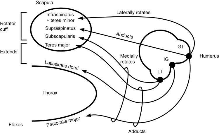
- The supraspinatus, not surprisingly (because it starts on top of the scapula), runs superiorly onto the lateral side of the humerus, where it inserts on the greater tubercle and abducts the arm.
- The deltoid runs in the same direction, but being superficial to the supraspinatus naturally extends farther down, inserting on the deltoid tuberosity, and also abducts the arm.
- The subscapularis, being on the anterior surface of the scapula, naturally inserts on the anterior bump of the humerus (= lesser tubercle), and medially rotates the arm.
- The infraspinatus and teres minor are close together and both wrap around posteriorly to insert on the greater tubercle, thus laterally rotate the arm.
- The teres major and latissimus dorsi both insert on the front of the humerus (teres major inserts at lesser tubercle; latissimus dorsi being the larger muscle, wraps around and extends further to the intertubercular groove). Thus, these both adduct, extend, and medially rotate the arm.
- Pectoralis major also "wants" to medially rotate the arm, but has to reach over all the other muscles on front of humerus (subscapularis, teres major, latissimus dorsi) to insert all the way on the greater tubercle. Actions of pectoralis major are similar to latissimus dorsi, except that pectoralis major, as you would expect from its anterior origin, flexes rather than extending the arm.
- The coracobrachialis ("armpit muscle"), as you might expect, inserts on the medial humerus, thus adducts the arm (it also flexes the arm).
- (2/24/08) The diagram below shows insertions and actions of muscles inserting on the proximal humerus (deltoid and coracobrachialis are omitted for simplicity). GT = greater tubercle, IG = intertubercular groove, LT = lesser tubercle. Note that the lines do not cross, and tend to space themselves out (e.g. teres major, latissimus dorsi, and pectoralis major all insert at different locations), which can help you remember the insertions.
Forearm and hand
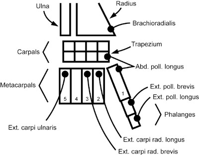
- (2/24/08) Insertions of posterior forearm muscles: There is logic to this arrangement, e.g.: brachioradialis inserts on radius; ext. carpi radialis longus inserts on radial edge of main part of hand (metacarpal 2); since that bone is "taken", the brevis inserts on metacarpal 3; ext. carpi ulnaris inserts on ulnar edge of hand (metacarpal 5). The three extrinsic thumb muscles each insert on a different bone, in the same order (proximal to distal) that the muscle bellies appear on the forearm.
- In forearm/hand, the "brevis" muscles of the thumb all insert on the proximal phalanx of the pollex:
- This includes extensor pollicis brevis, abductor pollicis brevis, flexor pollicis brevis.
- Of the intrinsic hand muscles for which OIA information is included in this course, all insert on the proximal phalanx of the pollex, except for the opponens pollicis, which inserts on the 1st metacarpal. This makes sense because inserting on this more proximal bone is better for performing opposition (medial rotation) of the thumb.
- Adductor pollicis: Origin: capitate bone and 2nd-4th metacarpals. There is in fact a close relationship between these bones. If you were to extend your 3rd digit among violent persons, you might risk being decapitated. This is a reminder that the 3rd metacarpal lies adjacent to the capitate bone. It is not too hard to remember that the metacarpals on either side (metacarpals 2 and 4) also participate in the origin of this muscle.
Anterior forearm
- Several muscles of the anterior (ventral) forearm originate on the medial epicondyle of the humerus. This makes sense because when relaxed, the ventral forearm tends to face medially.
Posterior forearm
- Several muscles of the posterior (dorsal) forearm originate on the lateral epicondyle of the humerus. This makes sense because when relaxed, the dorsal forearm tends to face laterally.
Hand and foot muscles compared
- (10/11/12) There are numerous similarities between the muscles of the thumb and little finger, and between the muscles in the palm of the hand and the sole of the foot. (Some of the forearm and leg muscles that have actions similar to the intrinsic muscles are shown as well.)
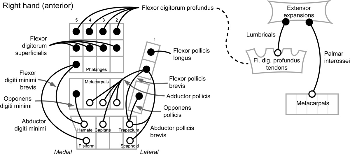
- The flexors of the thumb and little finger both attach to the proximal phalanx and the closest carpal.
- The opponens of the thumb and little finger both attach to the closest metacarpal and carpal; the action of these muscles (opposition) is to cup the hand, by pulling the metacarpal to the opposite side.
- It makes sense that the abductor muscles, which move at right angles to the flexor muscles, need a different origin (pisiform for little finger, scaphoid for thumb). You can argue the thumb abductor also needs an extra origin (trapezium) because it is larger than the little finger.
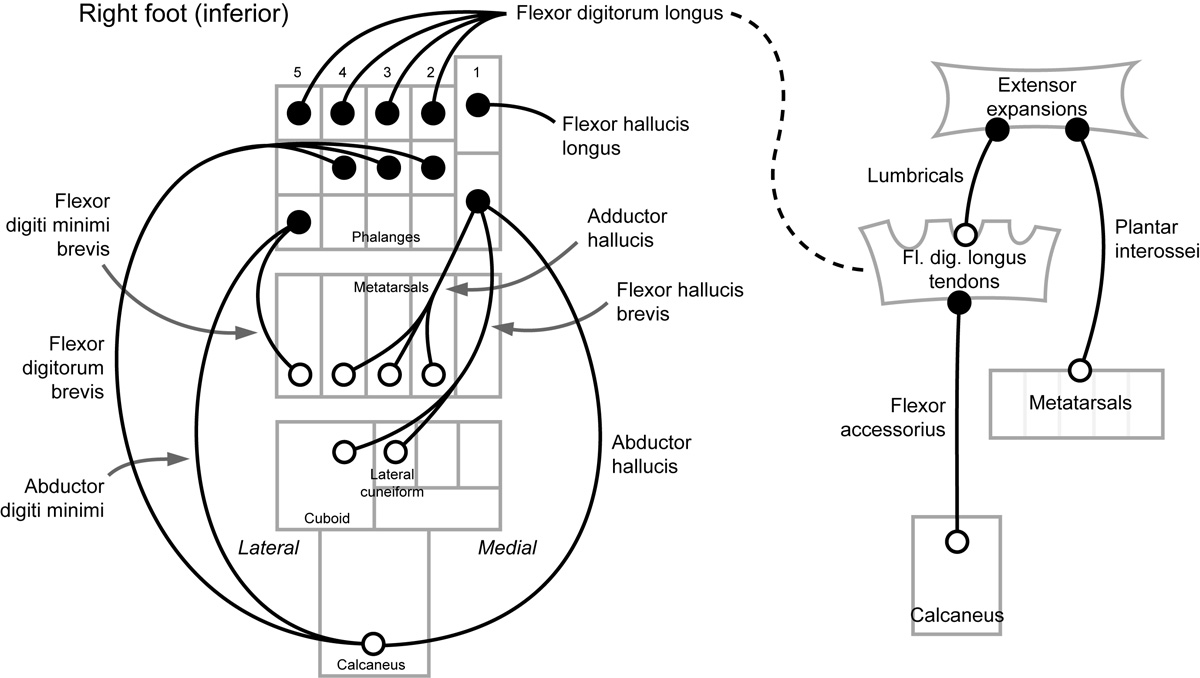
- The flexor hallucis longus and flexor pollicis longus both insert on the distal phalanx.
- In the first layer of foot muscles, the flexor digitorum brevis is comparable to the hand's flexor digitorum superficialis; they are both superficial and insert on the middle phalanges.
- The foot's two abductor muscles have similar origin and insertion.
- In the second layer of foot muscles, you find the tendons of the flexor digitorum longus, and muscles attached to it. The flexor digitorum longus is comparable to the hand's flexor digitorum profundus; they both insert on the distal phalanges, and are the origin for the lumbricals, which insert on extensor expansions (and have the same actions) in both hand and foot. (See typo note below.)
- In the third layer of foot muscles, the adductor hallucis has attachments similar to those of the hand's adductor pollicis.
- However, the flexor hallucis brevis, surprisingly (since the hallux is medial), has origins on the lateral side of the foot. The best way to remember this is that the flexor hallucis longus also has its origin, surprisingly, on the lateral side (of the leg).
- In the fourth layer, the plantar interossei have the same attachments as the hand's palmar interossei. These include the same actions as the lumbricals, but also adduction of the digits. (Typos: The 8th edition of the lab manual should say plantar interossei adduct, rather than abduct, the toes. Also, the foot lumbricals and plantar interossei actions should say "flex metacarpophalangeal joint, extend interphalangeal joints" like the lumbricals of the hand.)
Medial compartment of thigh
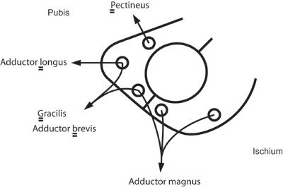
- (2/22/08) The four muscles of the medial compartment of the thigh, together with the pectineus (listed in anterior compartment), have related origins, insertions and actions, as discussed below.
- (2/22/08) Origins: See diagram to the right. For simplicity, the parts of the pubis and ischium are not labeled here. Note: The adductor magnus is a large muscle with several origins. The other muscles in the diagram go roughly in reverse alphabetical order (as indicated), from top to bottom. Note: lab manual says adductor longus originates on pubis near pubic symphysis, which is simplified here to body of pubis.
- (2/22/08) Insertions: Four of these muscles insert on the linea aspera of the femur. The long & graceful gracilis inserts all the way on the medial tibia, which explains the different actions it performs (see table of actions, below).
- (10/5/2011) Actions: All muscles in this group (medial compartment plus pectineus) perform "adduction, medial rotation, and flexion of the thigh" except: (1) Adductor brevis, being a short muscle, cannot do flexion and (2) Gracilis, because it crosses the knee, performs medial rotation and flexion on the leg, not the thigh.
Adduct Medially rotate Flex pectineus Yes Yes Yes gracilis Yes Yes (leg) Yes (leg) adductor longus Yes Yes Yes adductor brevis Yes Yes adductor magnus Yes Yes Yes
Gluteal muscles and posterior thigh muscles
- Actions of the gluteal muscles: The gluteus MEDius rotates the thigh MEDially. The gluteus mInimus likewise rotates the thigh medIally, which makes sense because these two muscles share the same approximate origin and insertion (as you can see on the cadavers as well). In contrast, the gluteus mAximus rotates the thigh lAterally.
Leg
- The following diagram shows the bones (and interosseous membrane) on which the leg muscles (other than soleus and gastrocnemius) originate. Note that this does not give all the details you need to know (e.g. lateral condyle, etc). T = tibia, I = interosseous membrane, F = fibula, TA = tibialis anterior, FB = fibularis brevis, FHL = flexor hallucis longus, etc.
- Draw a line from each of the 3 fibularis muscles to the fibula (this makes sense).
- Draw lines from the big toe muscles (EHL and FHL) to F and I. Mnemonic: "tap your toe to the music from the hiFI (or wiFI)".
- For all the muscles that still have no line (TA, EDL, TP, FDL), draw a line to T. (Makes sense since T is a big bone.)
- All anterior muscles connect to the interosseous membrane. (Add lines from TA, EDL, and FT to I.)
- The EDL and TP connect to all three origins, so add lines for these muscles. Mnemonic: "Ed'll T.P. your house all over", i.e. the EDL and TP muscles have lines going to all three origins.
- (10/4/2011) The following diagram shows the positions of the leg muscle origins on the leg.
- None of the lines indicating the muscles cross.
- The origins on the bones occur at roughly four heights: Head/condyle, proximal shaft, middle shaft, and distal shaft.
- All muscles of the anterior compartment have an origin on the interosseous membrane. Neither one of the lateral compartment muscles has an origin there.
- Counting each muscle's origins separately, the digitorum muscles together have 4 origins, the hallucis muscles together also have 4 origins, and the fibularis muscles together have 4 origins as well.
- Both hallucis muscles originate on fibula and interosseous membrane (see "wiFI" mnemonic above).
- All fibularis muscles originate on the fibula.
- See "Ed'll T.P. your house all over" mnemonic above.
- The fibularis longus, being a longer muscle, extends farther up the fibula than the brevis.
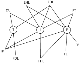
You can reconstruct this diagram with the following steps:
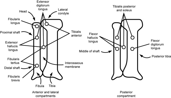
Notice that:
Intrinsic back muscles
- To remember the numbers of the vertebrae upon which these muscles originate or insert, two-word phrases are handy, as explained below.
- Iliocostalis: Origin: iliac crest, ribs 3-12. The iliac crest makes sense, given the name of the muscle. To remember the ribs, think "Rib R". The word "rib" has 3 letters, and the letter "R" when pulled apart looks like a "12". Hence "ribs 3-12". Insertion: inferior border of ribs, transverse processes of C4-C6. Insertion on ribs makes sense, given the name of the muscle ("costal" = "rib"). To remember C4-C6, think "cost cutter", i.e., a 4-letter word beginning with C, and a 6-letter word beginning with C.
- Longissimus: Origin: transverse processes of C4-L5. To remember this, think "crab lover" (or your favorite 4-letter word beginning with C, and a 5-letter word beginning with L). Insertion: mastoid process of temporal bone. This is logical, since this muscle is located in a lateral position on the back, and the mastoid process is in a lateral position as well.
- Spinalis: Origin: spinous processes of T7-L3. As you no doubt have learned, "I love spaghetti" is a mnemonic for the iliocostalis, longissimus, and spinalis muscles, respectively. Thus, a person over the age of 21 who is fond of Italian cuisine and beverages might choose to responsibly drink "iTalian aLe" with his/her spaghetti meal. This phrase consists of a 7-letter word with a T, and a 3-letter word with an L, hence T7-L3. Insertion: spinous processes of C4-T6. Think "cold turkey" (4-letter C word, followed by 6-letter T word), which might be a reasonable choice for someone who has consumed too much Italian ale. (Note: One definition of "cold turkey" is "immediate, complete withdrawal from something on which one has become dependent".)
- Splenius capitis: Origin: spinous processes of C6-T7. Think "spleen trouble". The 7-letter word "Trouble" indicates T7, and the 6-letter word "spleen" indicates C6 (even though "spleen" does not contain a C, "s" sounds a bit like "c"). After the muscle exam, you will learn about an organ in the upper left abdomen called the spleen. One of your splenius capitis muscles, in fact, points roughly in the direction of the spleen. Insertion: mastoid process of temporal bone, occipital bone. This is logical because this muscle inserts broadly at an angle across much of the back of the head, so it attaches to both lateral structures (the mastoid processes) and medial structures (the occipital bone).
- Semispinalis capitis: Origin: transverse processes of C7-T12. "Capitis" is a 7-letter word beginning with C. "Semispinalis" is a 12-letter word, so it has to represent T12, because C12 and L12 do not exist. Insertion: Occipital bone. This is logical, because this muscle lies right next to the midline and travels straight up the back, toward the occipital bone.
- Note on the vertebrae listed above: All of these refer to the transverse processes, except for the following muscles which attach to the spinous proceses: spinalis (makes sense, given the name of the muscle) and splenius capitis (makes sense when you remember that this muscle travels obliquely from the head toward the midline of the back).